

Modern eye care demands a higher level of diagnostic precision than ever before. With increasing screen exposure, lifestyle changes, and age-related ocular conditions, patients expect accurate diagnoses and dependable treatment outcomes. Clinical accuracy is no longer just about identifying obvious symptoms—it involves detecting subtle structural changes in the anterior segment of the eye before they progress into serious issues. Advanced anterior segment imaging technologies have transformed the way clinicians approach diagnosis, offering detailed visualization that significantly improves decision-making and patient outcomes.
The anterior segment, which includes the cornea, iris, anterior chamber, and lens, plays a crucial role in vision clarity. Even minor abnormalities in these structures can affect visual performance. Advanced imaging tools allow practitioners to identify micro-level changes that may not be visible through traditional examination techniques.
Understanding Anterior Segment Imaging
Anterior segment imaging refers to technologies that provide magnified, illuminated, and high-resolution views of the front structures of the eye. These systems combine precision optics with controlled light beams to highlight tissue layers and surface details. By producing sharp cross-sectional or surface images, clinicians can evaluate corneal health, assess lens clarity, and monitor inflammatory conditions more effectively.
The integration of digital imaging capabilities has elevated these tools beyond simple visualization. Today’s systems allow image capture, storage, and comparison over time. This longitudinal tracking improves clinical monitoring and ensures that treatment plans are based on measurable changes rather than subjective observation.
Early Detection of Subtle Ocular Changes
One of the most important advantages of advanced anterior segment imaging is early detection. Many eye conditions, such as keratitis, early cataract formation, or corneal dystrophies, begin with minor structural variations. Without magnified and controlled illumination, these changes can be overlooked.
High-quality imaging provides enhanced contrast and depth perception, allowing practitioners to detect small epithelial defects, micro-deposits, or early inflammatory signs. Early detection leads to timely intervention, reducing the risk of complications and improving long-term visual outcomes.
By identifying problems at their earliest stage, clinicians can recommend preventive measures or initiate targeted treatment, ultimately protecting patients from progressive vision loss.
Improved Accuracy in Diagnosis and Treatment Planning
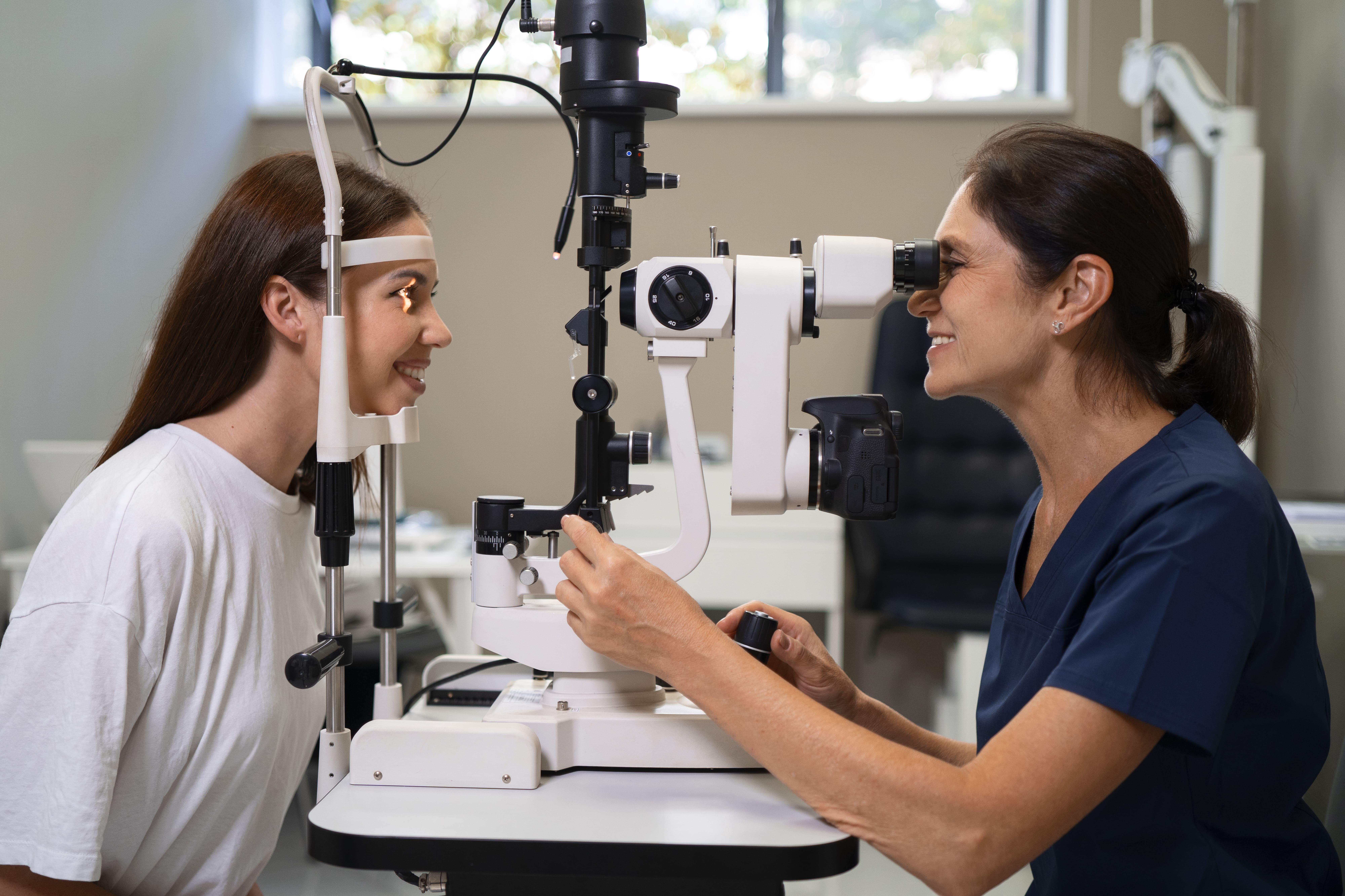
Accurate diagnosis forms the foundation of effective treatment. Advanced imaging tools support detailed evaluation of ocular structures, minimizing guesswork during examination. The use of a precision-based system like a slit lamp enables clinicians to carefully assess corneal transparency, anterior chamber depth, and lens clarity with remarkable detail.
This level of clarity helps differentiate between similar-looking conditions. For example, distinguishing between various types of conjunctivitis or identifying specific corneal ulcers becomes more reliable when supported by enhanced magnification and illumination. As a result, treatment plans become more accurate and personalized.
Furthermore, precise imaging assists in evaluating the effectiveness of ongoing therapies. By comparing images over multiple visits, clinicians can determine whether medications or interventions are producing measurable improvements.
Enhanced Documentation and Clinical Confidence
Digital imaging capabilities allow clinicians to capture high-resolution photographs of the anterior segment. These images serve as valuable documentation for medical records and can be shared with patients for better understanding of their condition.
Clear documentation strengthens clinical confidence by providing visual evidence to support diagnostic decisions. It also facilitates collaboration between specialists when referrals are required. For instance, if a patient needs advanced surgical evaluation, detailed images offer a clear starting point for further assessment.
Additionally, visual documentation enhances patient communication. When patients see the actual condition of their eye structures, they are more likely to understand the need for recommended treatments or follow-up visits.
Supporting Safer and More Effective Procedures
Advanced anterior imaging not only improves diagnostics but also enhances procedural safety. Before performing procedures such as foreign body removal or minor anterior segment surgeries, clinicians rely on precise visualization to ensure accurate execution.
Detailed imaging allows for careful evaluation of corneal thickness, lesion location, and tissue response. This reduces the risk of procedural errors and improves surgical outcomes. By offering a clear and magnified field of view, modern imaging tools help practitioners work with greater precision and confidence.
Streamlining Workflow in Busy Practices
Efficiency is essential in modern clinical environments. Advanced imaging systems are designed to provide quick alignment, stable positioning, and easy focus adjustments. This reduces examination time while maintaining high diagnostic standards.
Faster image capture and digital storage eliminate the need for repetitive examinations. Instead of relying solely on manual notes, clinicians can review captured images instantly. This not only saves time but also ensures consistency across multiple visits.
Streamlined workflow benefits both practitioners and patients. Reduced waiting times and quicker consultations enhance overall patient satisfaction without compromising diagnostic quality.
Elevating the Standard of Patient Care
Ultimately, advanced anterior segment imaging raises the overall standard of eye care. By combining magnification, illumination, and digital analysis, these technologies empower clinicians to make data-driven decisions.
Patients benefit from earlier diagnosis, more accurate treatment plans, and improved communication. Whether managing routine eye conditions or complex anterior segment disorders, precise imaging ensures that care is based on reliable visual evidence.
As technology continues to evolve, anterior segment imaging systems will likely incorporate even more sophisticated features such as enhanced digital analytics and integration with electronic medical records. These advancements will further strengthen clinical accuracy and elevate the quality of vision care services.
Conclusion
Advanced anterior segment imaging has become a cornerstone of modern ophthalmic practice. By enabling early detection, improving diagnostic precision, supporting effective treatment planning, and enhancing clinical documentation, these technologies significantly improve clinical accuracy.
In today’s patient-centered healthcare environment, precision is not optional—it is essential. With the support of advanced imaging systems, clinicians can deliver safer, more reliable, and more efficient eye care. As practices continue to adopt innovative diagnostic tools, the future of anterior segment evaluation looks increasingly accurate, efficient, and patient-focused.